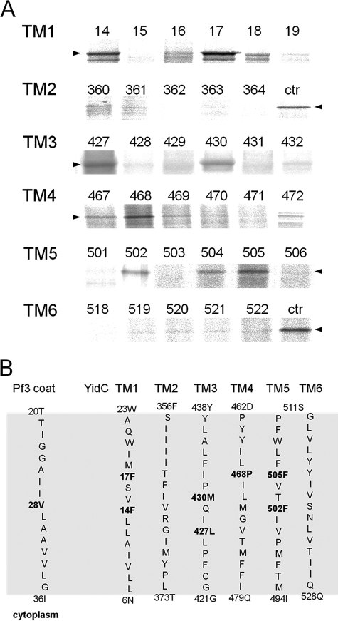
FIGURE 2.
Disulfide complexes of inserting Pf3-28C with YidC cysteine mutants in the center of TM1 to TM6. A, MK6 cells bearing the plasmid pMS-Pf3-28C and a second plasmid, pACYC, encoding the respective single cysteine YidC mutant, were grown in the presence of 0.2% glucose to deplete the cells from the chromosomally encoded YidC. The cells were induced with 1 mm IPTG for 10 min and pulse-labeled with [35S]methionine for 30 s. The cells were put on ice and treated with 1 mm copper phenanthroline. The cells were TCA-precipitated, immunoprecipitated with antibody to the Pf3 virus, and analyzed on non-reducing SDS-PAGE by phosphor imaging. For the control lanes, Pf3-28C and YidC430C (ctr) were coexpressed and treated as described above. The arrowheads depict the position of YidC-Pf3 complex. B, positions of the Pf3-28C contacts with YidC in the six transmembrane segments. The residues that provided stable disulfide cross-links when mutated to a cysteine residue are shown in boldface. The contacts were found in the center of TM1, 3, 4, and 5.

