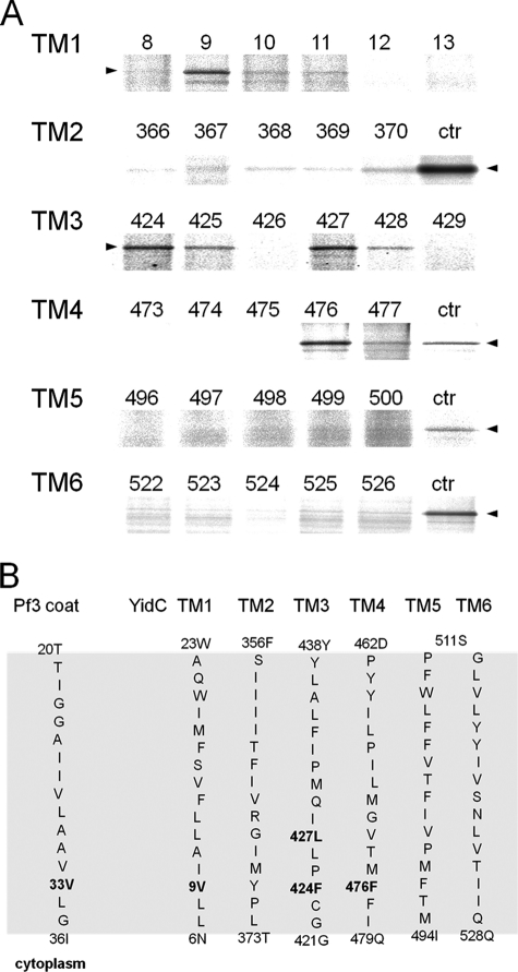
FIGURE 4.
Inserting the Pf3-33C coat protein contacts YidC transmembrane residues located in the inner leaflet. A, E. coli MK6 cells bearing the plasmid pMS-Pf3-33C and a second plasmid encoding the respective single cysteine YidC mutant were grown and treated as described in the legend to Fig. 2. Cross-links were detected by immunoprecipitations with an antibody to Pf3. For the control lanes, Pf3-28C and YidC505C (ctr) were coexpressed and treated as described above. The arrowheads depict the position of the YidC-Pf3 complex. B, positions of the Pf3-33C contacts with YidC in the six transmembrane segments. The residues that provided stable disulfide cross-links when mutated to a cysteine residue are shown in boldface. The contacts were found in the inner half of TM1, 3, and 4.

