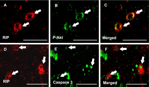
FIGURE 10.
Akt and caspase 3 are activated in OLs in vivo. Confocal micrographs show immunofluorescent co-localization using the OL-specific antibody anti-RIP (A and D) in combination with antibodies generated against phosphorylated Akt (Ser-473) (B, MCAO sham-operated vehicle treated rat) or activated caspase 3 (E, MCAO vehicle-treated rat) in the external capsule. Merged images demonstrate expression of phosphorylated Akt (C) and activated caspase 3 (F) in RIP-positive OLs. Arrows denote immunoreactivity. Scale bars = 50 μm.

