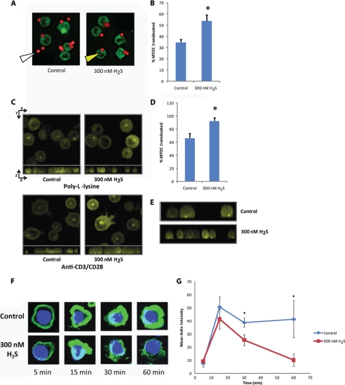
FIGURE 4.
H2S targets the cytoskeleton in activated T cells. A, anti-CD3/CD28-coated magnetic beads (red) were incubated with EYFP-α-tubulin expressing Jurkat cells (pseudo-colored green) in the presence or absence of 300 nm Na2S for 15 min at 20% O2. Yellow arrow = MTOC; white arrow = microsphere. B, MTOC orientation to the anti-CD3/CD28-coated magnetic beads was quantified as % translocated. n = 50 cells for each condition. *, denotes p < 0.05. C, EYFP-α-tubulin expressing Jurkat cells (25,000) were added to either poly-l-lysine or anti-CD3/CD28-coated glass coverslips with or without 300 nm Na2S and incubated for 45 min before confocal images of z-stacks were taken. At the top of each panel is the composite image of the x,y plane and directly under is the x,z plane. D, MTOC orientation to the center of the immunological synapse created at the glass surface was quantified as % translocated. n > 25 cells for each condition. * denotes p < 0.05. E, shown are images from the x,z plane anti-CD3/CD28-coated portion of D cropped to a single cell depth. F, the actin cytoskeleton (green) of fixed Jurkat cells was stained with phalloidin after stimulation with anti-CD3/CD28 in the presence or absence of 300 nm Na2S for the denoted times and imaged using confocal microscopy. Images are maximum projection composites of Z-stacks. G, quantified intensity of actin staining at each time point is shown.

