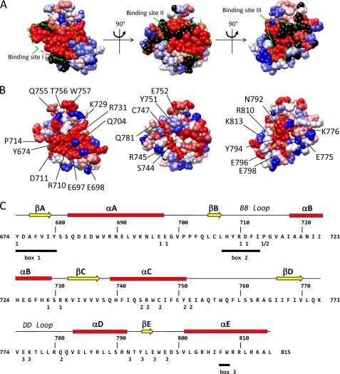
FIGURE 2.
Identification of three candidate binding sites in a homology model of the TLR4 TIR domain. The model is shown in three orientations. The suggested candidate binding sites are indicated by a dashed line. A, residue conservation of TLR4. Residues are colored according to the ClustalX (45) score in an alignment of 29 TLR4 orthologues. Red, highly conserved; blue, less conserved. B, indication of the alanine scanning mutagenesis data of Ronni et al. (25). Residues are colored according to the NF-κB signal versus the WT in that study. Blue, 100% of WT; red, 0% of WT; black, not mutated in that study. C, secondary structure elements, box 1–3 motifs and position of binding sites I–III in the human TLR4 sequence. Binding sites I–III residues are labeled 1–3 below the sequence. The box 1–3 motifs of the TIR domain as originally defined by Slack et al. (46) are underlined.

