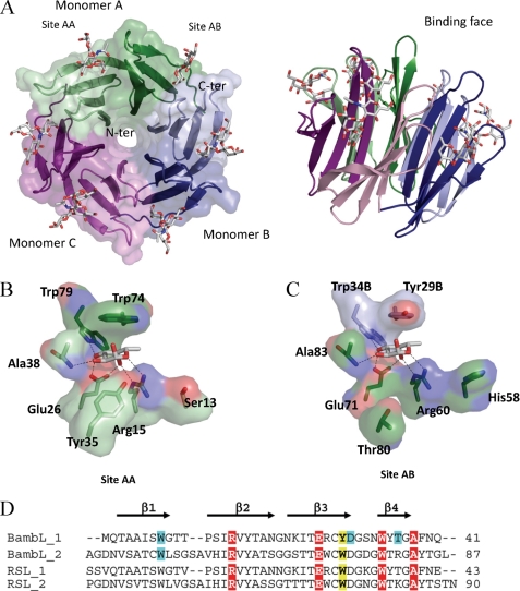
FIGURE 3.
Crystal structure of BambL/H-type 1 oligosaccharide. A, two representations of the six-bladed β-propeller formed by the association of three monomers, which are colored in green, blue, and purple. B and C, details of the interaction between fucose and BambL lectin in the intramolecular and intermolecular binding sites, respectively. Amino acids are from chain A unless indicated otherwise. D, sequence alignments of the two-blade repeat of BambL and comparison with RSL. Amino acids involved in hydrogen bonds and van der Waals contact with fucose have a red and yellow background, respectively. The ones involved in hydrogen bonds with other residues of the oligosaccharides have a blue background.

