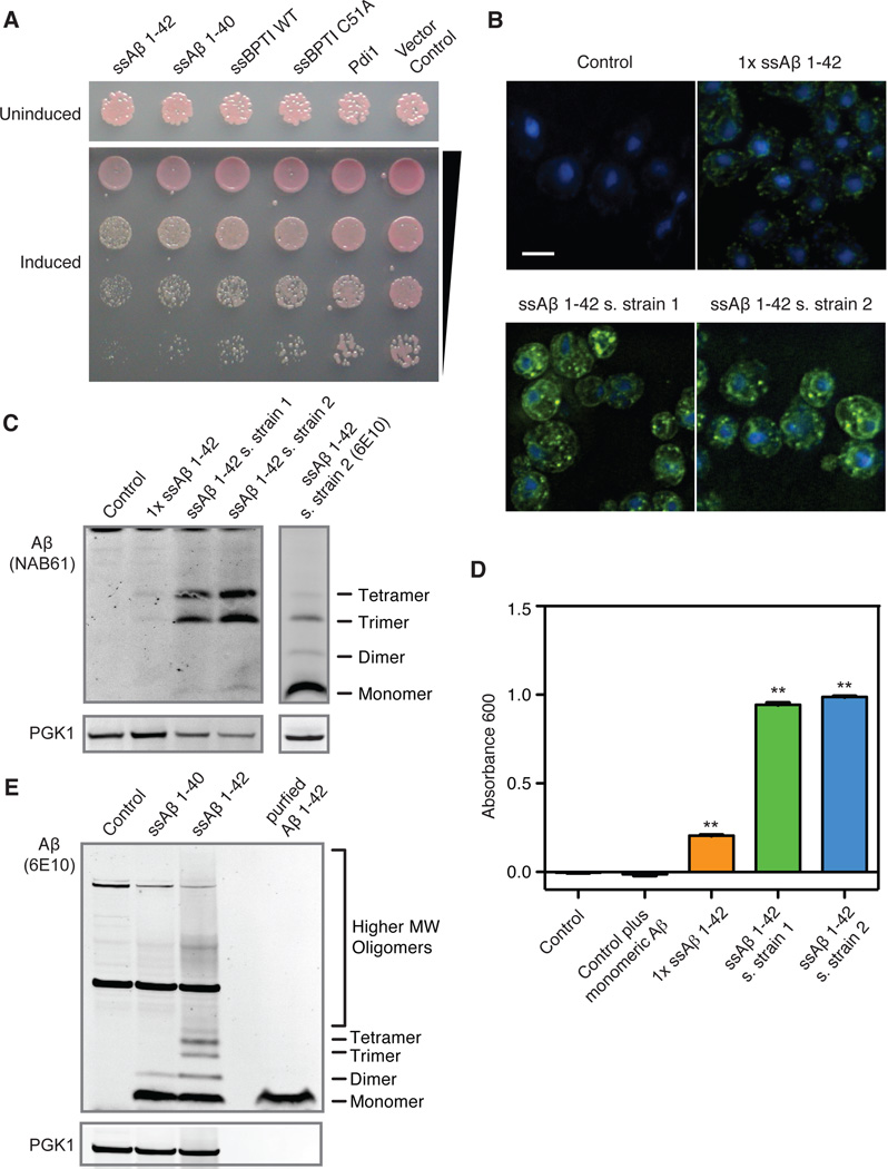
Figure 1.
Expression of Aβ in the yeast secretory pathway. (A) Comparison of ssAβ 1-42 toxicity with ssAβ 1-40, ssBPTI (WT and C51A) and Pdi1. Proteins were expressed using the inducible GAL1 promoter and a high copy number plasmid. Strains carrying the plasmids were serially diluted and spotted on inducing (galactose) and non-inducing (glucose) media. (B) Immunostaining for ssAβ 1-42 reveals localization to the ER/secretory pathway. Aβ was detected in the ER (ring surrounding the nucleus, stained blue with DAPI) and in small foci throughout the cell. The scale bar is 5µm. All figures are on the same scale. (C) Immunoblot with NAB61, an antibody specific for soluble Aβ oligomers, detects oligomers in unboiled samples. An immunoblot with the 6E10 Aβ-specific antibody is shown for reference. (D) An indirect ELISA assay using a monoclonal Aβ oligomer-specific antibody detects Aβ oligomers in the 1×ssAβ strain and, more so, in the two screening strains (n=5; error bars to small to be visible; **: p<0.01, based on Dunnett’s test). (E) Aβ 1-40 and Aβ 1-42 expression was detected by immunoblot analysis of unboiled lysates using 6E10.

