
Figure 4.
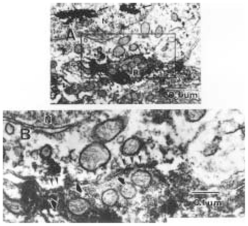
(A) A cholera toxin β subunit (CTβ)-immunoreactive (ir) retinal terminal synapses on the perikaryal membrane of a Calbindin-D28K-ir cell, indicated by the presence of tetramethylbenzidine crystals in the nucleus (arrows). Numerous mitochondria are observed on both the presynaptic and postsynaptic sides of the synaptic profile (magnification = × 34,000). At higher magnification (B), two postsynaptic enlargements (arrow heads) are visible near each other. Synaptic vesicles (black/white arrows) are present in the presynaptic profile near each synapse, although they are partially obscured by the intense immunolabel. Magnification = × 97,000.