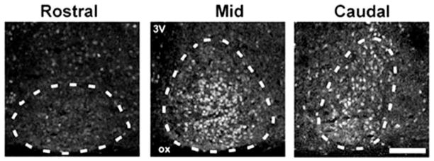
Fig. 2.
Photomicrographs depicting AR-ir in coronal sections in rostral (left), mid (center), and caudal (right) aspects of the SCN showing that AR-ir cells are concentrated within the mid and caudal aspects of the SCN. Dotted lines delineate the extent of the SCN as defined by AVP double labeling (not shown). Scale bar, 150 μm. ox, Optic chiasm; 3V, third ventricle.

