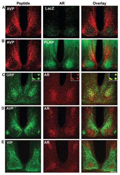
Fig. 3.
A and B, Photomicrographs depicting lacZ and PLAP reporters in the mid-SCN of AR-transgenic mice. The left column shows AVP, the middle column shows an AR reporter, and the right column shows the overlay. AVP (red) is used as a marker for the extent of the SCN. LacZ (A, green) staining confirms the results in Fig. 2, indicating that AR cells are highly concentrated in the ventrolateral core SCN. PLAP staining (B, green) also confirms the previous results and shows fibers of AR-containing cells fill the core SCN and are less dense in the dorsomedial shell region delineated by the AVP staining. There is a dense fiber plexus within the SCN, and efferents appear to exit dorsally from the nucleus. The area of intense PLAP staining dorsal to the SCN are AR fibers in the bed nucleus of the stria terminalis. C–E, Colocalization studies made use of double-labeled immunofluorescence in 35-μm coronal sections, stained for GRP (C), AVP (D), and VIP (E). The left column shows the peptide, the middle column shows AR, and the right column shows the overlay. Scale bar, 150 μm. ox, Optic chiasm; 3V, third ventricle. Inset on upper right of each panel in upper row shows a high-powered image from a 1-μm confocal scan.

