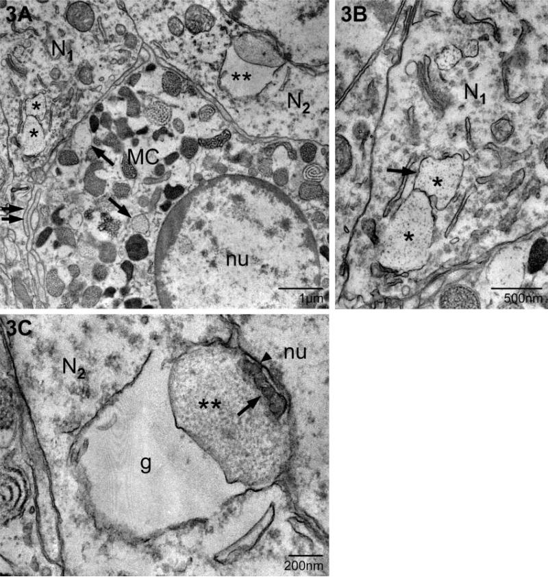
Fig. 3.
Localization of mast cell granule remnants in neighboring neurons. (A) Low magnification of a mast cell (MC) and adjacent neurons. Note the heterogeneity in size, shape and electron density of the granules that fill the mast cell cytoplasm. The arrows indicate electron-lucent, particle-filled granules characteristic of ongoing or recovery from exocytosis. Two neurons (N1 and N2; as shown in Fig. 1) contain mast cell granules (* and **, respectively). (B) A higher magnification of the granule remnants in A designated with *. In this section, two of the four contiguous remnants (*) (as identified in a set of serial sections, not shown) are seen. The granular material is enclosed by membrane (arrow). (C) A higher magnification of the granule remnant (**) in N2. This section is separated from that shown in A by ~200 nm. The remnant is composed of two chambers, one swollen and devoid of particles (g) and a compartment which retains particulate (**) and vesicular substructure (arrow). The second compartment is in contact with the outer nuclear envelope (arrowhead). nu, nucleus.

