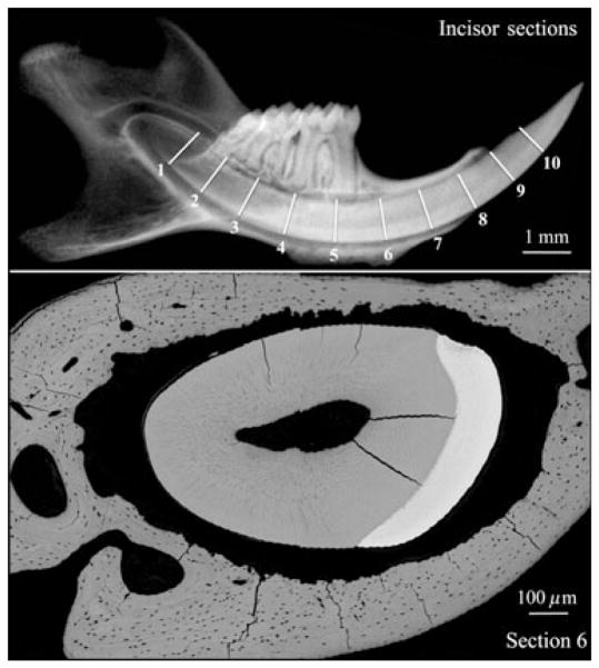
Fig. 2.
Radiograph (upper image) showing the approximate positions of incisor cross-sections on a mouse mandible at 9 wk, and a backscatter scanning electron microscopy image (lower image) of a cross-section sliced at position 6. In Fig. 3 we show incisor backscatter scanning electron microscopy images that we estimate to be from sections 2, 4, and 6 shown in the upper image of this figure, although the level of precision might have varied by as much as 1 mm.

