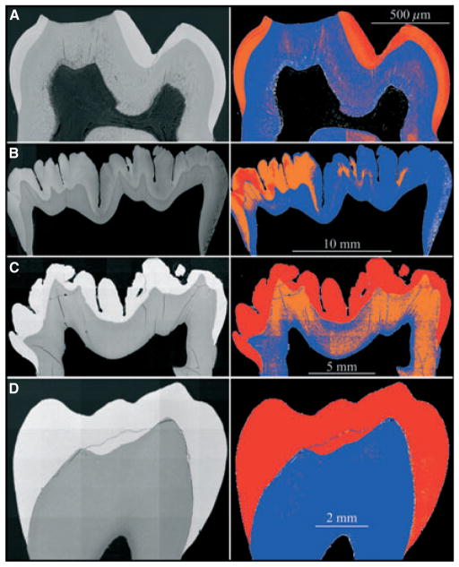
Fig. 6.
Backscatter scanning electron microscopy images of mouse, pig, and human molars. (A) First molar from 9-wk-old mouse. Enamel maturation is too advanced to detect a hypermineralized line at the dentino–enamel junction (DEJ). The maturation-stage crown is slightly more mineralized at the surface compared with the deeper regions of the enamel near the dentin. (B) Second molar from a 6-month-old pig. This unerupted molar has just finished the crown-formation stage and is starting to form roots. One side has a more advanced level of mineralization within the enamel layer. A hypermineralized line is not evident in the colour image but can be distinguished covering dentin near the cervical margin on the left. (C) First molar from a 6-month-old pig. This erupted molar is mature beyond the point where a hypermineralized line along the DEJ can be observed. (D) Premolar from a 14-yr-old human. The enamel layer is fully mature. The backscatter images were normalized to have the same mean gray-level intensity as that of the alveolar bone in the 9-wk-old wild-type mouse (Fig. 3).

