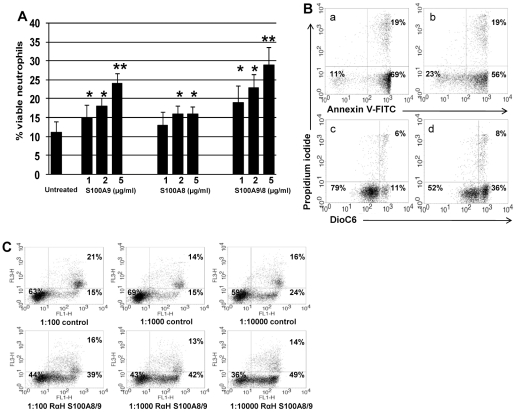
Figure 4. Effect of S100A9 and S100A8 on neutrophil apoptosis.
A. S100A9 and S100A8 add-in experiments. Varying concentrations of S100A9, S100A8, and the S100A8/9 complex were added to supernatants of neutrophils undergoing spontaneous constitutive PCD at a concentration of 0.5×10⁶/ml. After 10 hours apoptosis was evaluated using Annexin V-PI and mitochondrial staining. Data is presented as mean ± SD of Annexin V-PI staining (* p<0.05, ** p<0.02). B. S100A9 and S100A8 add-in experiments. A representative sample of Annexin V-PI and mitochondrial staining. Neutrophils undergoing spontaneous constitutive PCD without (a and c) or with (b and d) the addition of 2 µg/ml S100A8/9. Cells were stained either with Annexin V-FITC (a and b) or with DiOC6 (c and d), as well as PI (a, b, c, d) and assessed by flow cytometry. Dot plots are representative of 6 experiments. C. The effect of anti-S100A8 and A9 on add-in experiments. Neutrophils undergoing spontaneous constitutive PCD at a concentration of 0.5×10⁶/ml for 12h, with rabbit polyclonal antibody dilutions of 1∶100, 1∶1000, and 1∶10,000 against S100A9 and S100A8, were then treated with 2 µg/ml S100A8/9 complex. Rabbit serum at the same dilutions of 1∶100, 1∶1000, and 1∶10,000 was used as a control. Apoptosis was assessed using Annexin V-FITC and PI staining. Percentages of viable, early-, and late apoptotic cells are indicated within the respective quadrants. Dot plots are representative of 6 experiments.

