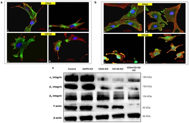
Figure 6. Expression of F-actin and integrins and their co-localisation with CD44/CD155 on GBM cells.
a, Co-staining of F-actin (green/left panels/L) or β1-integrin (green/right panels/R) with CD44 (red/top panels) and CD155 (red/bottom panels) on wild type UPAB cells. b, Co-staining of αvβ1-integrin (green/L) or αvβ3-integrin (green/R) with CD44 (red/top panels) and CD155 (red/bottom panels) on wild type UPMC (CD44 staining) and non-targeting siRNA treated SNB-19 cells (CD155 staining). All images in a and b have scale bars of 25 µm. c, Western blotting showed reduced expression of F-actin and integrins (αv, β1 and β3) in CD44/CD155-KD SNB-19 cells when compared to non-targeting siRNA treated cells (control).

