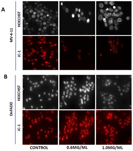
Figure 6. DRE destabilizes the mitochondria membrane potential of MV-4-11 cells.
(a). Following treatment with DRE, MV-4-11 cells were incubated with JC-1 dye to detect the loss of mitochondrial potential (ref to Materials and Methods). Red fluorescence indicates only cells that have healthy mitochondria. The mitochondria of MV-4-11 cells are completely destabilized by DRE treatment. On the other hand, the mitochondria of DnFADD cells remained unaffected by DRE treatment (b). Magnification: 400×.

