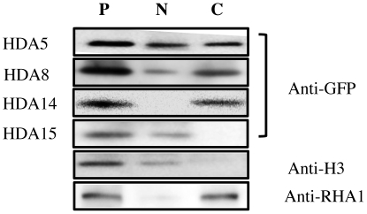
Figure 3. Cell fractionation and immunoblot detection HDA-GFP transfected protoplasts were separated into cytoplasmic and nuclear fractions then subjected to immunoblot analysis using anti-GFP antibody.
Histone H3 and RHA1 were used as nuclear and cytoplasmic markers, respectively, on WT protoplasts. P protoplast extract, N nuclear fraction, C cytosolic fraction.

