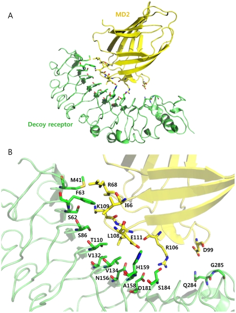
Figure 1. Identification of the mutation sites on the decoy receptor, TV3, for constructing single variants.
(A) Crystal structure of the decoy receptor in complex with MD2 (PDB ID 2Z65). The decoy receptor and MD2 are shown as green and yellow, respectively. (B) Structure of the interaction interface. 14 identified residues in TV3 are indicated in green, and potential interaction residues in MD2 are represented in yellow.

