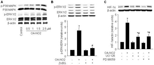
Figure 4. OA-NO2-induced HIF-1α is partially dependent on ERK1/2.
A) BAECs were incubated with the indicated concentrations of OA-NO2 for 16 h. Protein expression and phosphorylation of p38 MAPK and ERK1/2 were assayed as described in Materials and Methods. The blot is representative of three blots obtained from three independent experiments. B) BAECs were treated with ZnBG (1 µM) for 30 min followed by incubation with OA-NO2 for 16 h. ERK1/2 phosphorylation levels were monitored by immunoblot analysis, and band density was normalized to total ERK1/2 levels (lower panel). The blot is representative of three blots obtained from three independent experiments. *p<0.05 vs. control; # p<0.05 vs. OA-NO2 group. C) BAECs were treated with OA-NO2 alone or with the ERK1/2 signaling inhibitors, UO126 (10 µM) or PD98059 (50 µM), 1 h before the addition of OA-NO2. HIF-1α protein levels were monitored by immunoblot analysis, and band density was normalized to β-actin levels (lower panel). The blot is representative of three blots obtained from three independent experiments. *p<0.05 vs. control; # p<0.05 vs. OA-NO2 group.

