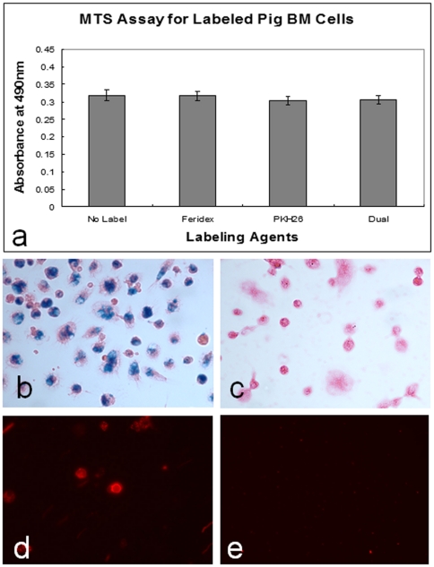
Figure 1. Results of in vitro studies.
(a) MTS assay shows the average proliferation efficiencies are almost the same among the different groups of BM-SPCs labeled with Feridex, PKH26, and dual agents (Feridex+PKH26), in comparison with control BM-SPCs without labeling. (b&c) Prussian-blue staining shows Feridex-positive cells as blue color spots, which are not seen in the unlabeled (control) cells (c). (d&e) Fluorescent microscopy demonstrates PKH26-positive BM-SPCs as red/orange-colored fluorescent dots (d), which are not visualized in unlabeled (control) BM-SPCs (e).

