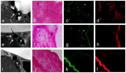
Figure 3. Representative MRI and corresponding histologic findings.
(a–d) Animal group 1 with Feridex/PKH26-dual-labeled cell transplantation, showing MR signal void of the iliac artery wall (arrows on a), Prussian blue-positive cells (b), dextran-positive cells (c), and PKH26-positive cells (d), as blue and green as well as red spots or dots, respectively, in the artery wall. (e–h) Animal group 2 with Feridex-only-labeled cell transplantation, demonstrating MR signal void of the iliac artery wall (arrows on e), Prussian blue-positive cells (f), dextran-positive cells (g) and a negative PKH26 stain (h). (i–l) Animal group 3 (control group) with unlabeled cell transplantation or no cell transplantation, showing the intact artery wall on MRI (arrow on i), a negative Prussian blue stain (j), a negative dextran stain (k), and a negative PKH26 stain (l). V = iliac vein.

