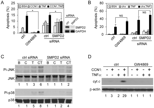
Figure 6. nSMase mediates CCN1/TNFα-, but not CHX/TNFα-induced apoptosis.
A, HSFs were either treated with 0.1% DMSO (ctrl) or 20 µM GW4869 for 30 mins, or subjected to siRNA-mediated silencing of nSMase1 (SMPD2). Cells were then tested for apoptosis in response to CCN1 (2 µg/ml) and/or TNFα (10 ng/ml). (*p<0.05; n = 3). On the right is RT-PCR analysis of SMPD2 and GAPDH expression in cells transfected with control or SMPD2 siRNA. B. Apoptotic response of cells to CHX (1 µg/ml) and/or TNFα was assessed after the inhibition of nSMase with GW4869, or upon siRNA-mediated silencing of nSMase1 (NS-not significant, n = 3). C, HSFs were transfected with control siRNA or siRNA against nSMase 1, and stimulated for 5 hrs with BSA (B), CCN1 (C), TNFα (T), or both (CT). Phosphorylation of JNK (Thr183/Tyr185) and p38 MAPK (Thr-180/Tyr-182) was analyzed by immunoblotting. Numbers under the immunoblots are relative signal intensities as determined by densitometry. D, HSFs were incubated with GW4869 (20 µM) where indicated, and treated with CCN1 and/or TNFα for 5 hrs. Cytosolic extracts were electrophoresed and cytochrome c was detected by immunoblotting. Numbers under the immunoblots are relative cytochrome c signal intensities as determined by densitometry.

