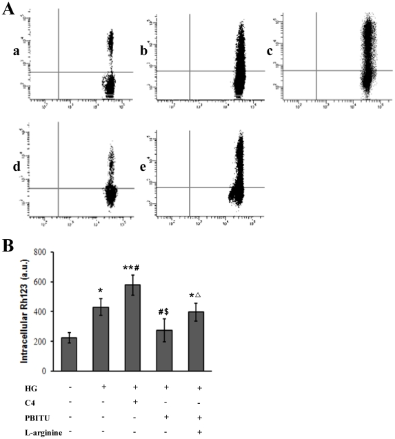
Figure 5. Decreased P-gp functional activity by high glucose involves iNOS.
(A) P-gp function was determined by intracellular rhodamine 123 accumulation by flow cytometry. D407 cells were incubated in normal (a) and high glucose medium (b–e), respectively. Cells incubated in high glucose were pretreated with C-4 (c), or PBITU alone (d) or combined with L-arginine (e) for 1 h. Following the treatment, cells were then loaded with a rhodamine 123 (10 µg/mL). The mean fluorescence intensity of intracellular rhodamine 123 was determined by flow cytometry. The results shown are representative of those for three separate experiments. (B) The bar represents the mean ± SE for the three separate experiments. *P<0.05 and **P<0.01 compared with cells incubated in normal glucose medium; # P<0.05 compared with cells incubated in high glucose; $ P<0.05 compared with cells incubated in high glucose and C4; Δ P<0.05 compared with cells incubated in high glucose and PBITU. HG, high glucose.

