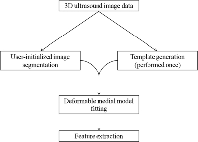
Figure 1.
Schematic of semi-automated image analysis. First, a deformable medial template of the mitral valve is generated from 3D ultrasound image data. This template generation step is performed once. Then, for each subject in the study, a 3D ultrasound image of the mitral valve is segmented using the level set method. The template is then deformed to fit each segmented image. Finally, morphological features are automatically computed from the fitted medial representation of each valve.

