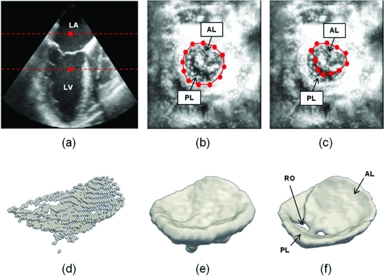
Figure 2.
Outline of semi-automated segmentation. (a) The user initializes two points in a long-axis cross-section of the 3D US image volume, identifying an ROI (red) containing the valve along the axial dimension. (b) The user initializes a series of annular points in an enhanced projection image depicting the valve from an atrial perspective. (c) The user shifts posterior annular points into the coaptation zone, forming an outline of the anterior leaflet in the enhanced projection image. (d) A 3D point cloud delineating the valve is automatically generated. (e) The 3D point cloud is morphologically dilated with a spherical structuring element to obtain an ROI containing the valve. (f) A final segmentation of the valve is obtained by thresholding and active contour evolution. (LA = left atrium, LV = left ventricle, AL = anterior leaflet, PL = posterior leaflet, RO = regurgitant orifice).

