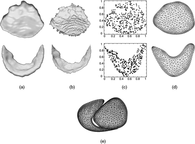
Figure 4.
Schematic of the template generation process. (a) A 3D US image volume is segmented to obtain binary images of the anterior (top) and posterior (bottom) mitral leaflets. (b) The binary images are skeletonized in 3D. (c) A 2D scatter plot of the skeletons’ vertices in u1,u2 space. (d) The constrained conforming Delaunay triangulation of the region containing the skeletons’ vertices. (e) The combined medial manifolds of the anterior and posterior leaflets used for deformable modeling.

