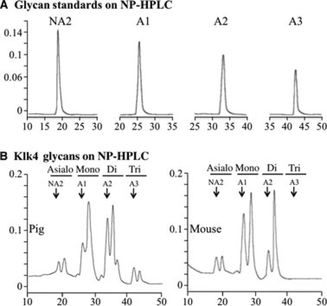
Fig. 2.
Sialidation of N-glycans. The y-axes represent fluorescence emitted at 420 nm and the x-axes represent minutes from injection of sample. (A) Normal-phase high-performance liquid chromatography (NP-HPLC) chromatograms of glycan standards containing no (NA2), one (A1), two (A2) and three (A3) sialic acid attachments.(B) NP-HPLC chromatograms of labeled N-glycans released from pig (left) or mouse (right) kallikrein-related peptidase 4 (KLK4). Arrows point to the retention times of the standards. Note that the N-glycans released from mouse KLK4 do not show peaks at retention times for three sialic acids.

