
Figure 5. Cys8 residue of cellular ANXA2 is a redox sensitive cysteine.
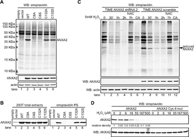
(A) 293T cells were transiently transfected with pcDNA3 empty vector (lane 1), pcDNA3-ANXA2 WT (lane 2), or a series of ANXA2 cysteine mutant plasmids: pcDNA3-ANXA2-Cys-8-Ala (lane 3), pcDNA3-ANXA2-Cys-8-Ser (lane 4), pcDNA3-ANXA2-Cys-132-Ala (lane 5) or pcDNA3-ANXA2-Cys-132-Ser (lane 6) for 48 hours. 20 μg of each cell extract was labeled with 20 μM BIAM and subjected to SDS-PAGE followed by western blotting with a streptavidin probe and the antibodies indicated. (B) 200 μg of each cell lysate was incubated with 20 μM BIAM, labeled proteins were purified with streptavidin beads. Cell extracts and streptavidin purified samples (streptavidin PS) were subjected to SDS-PAGE followed by western blotting for ANXA2. (C) TIME ANXA2 shRNA2 or TIME ANXA2 scramble cells were treated with 5 mM H2O2 in the absence or presence of 10 mM NAC for the times indicated. CA stands for competition assay; these samples were treated with 5 mM DTT prior to incubation with BIAM. Cell extracts were labeled with 10 μM BIAM, subjected to SDS-PAGE followed by western blotting with the antibodies indicated. (D) 0.65 μM of human recombinant ANXA2 or ANXA2C8S protein was incubated with the indicated concentrations of H2O2 for 30 minutes, after what 10 μg/ml of catalase was added. Samples were labeled with 20 μM BIAM and subjected to SDS-PAGE followed by western blotting with a streptavidin probe and ANXA2 antibody. Protein band quantification was done using the Licor Odyssey software.