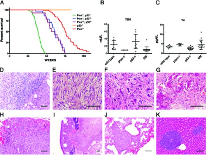
Figure 1. Clinicopathological features of [Pten, p53]thyr−/− mice.

(A) Kaplan-Meyer analysis of the effect of progressive p53 deletion on the survival of Ptenthyr−/− mice. (B, C) TSH and T4 serum levels in control, single mutant and double mutant (DM) mice. (D-K) Histopatological features of tumors developing in [Pten, p53]thyr−/− mice. (D) Anaplastic carcinoma (left) flanking an area of well-differentiated follicular carcinoma (right). Undifferentiated tumors display areas of spindle cell morphology (E), with frequent giant cells (F), and occasional bone metaplasia (G). Tumors invade locally into the muscle (H), and the trachea (I), and metastasize to the lungs (J) or, sporadically, to the liver (K). Bar: 100μm.
