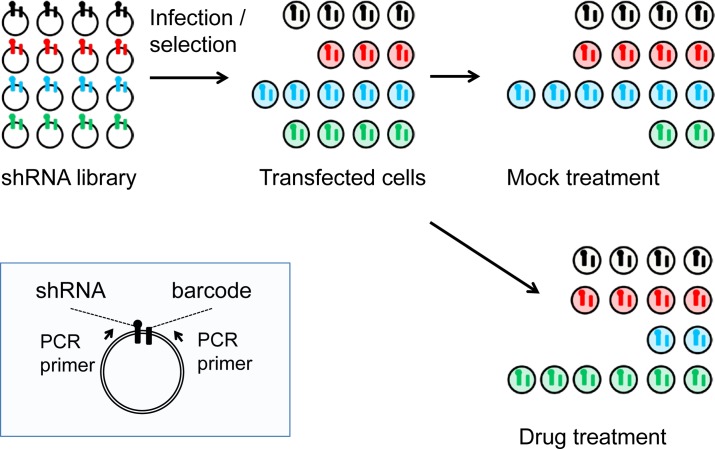
Figure 1. Schematic of the pooled shRNA screening methodology.
Shown in the blue box insert are features of the MLP retroviral vector including the two PCR primers that are used to amplify the shRNA and its linked barcode. Shown in the upper left is a representation of the relative abundance of individual shRNAs in the library as it is prepared in E. coli. Following transfection into mammalian cells and selection for stable integration, the relative abundance of individual shRNA changes, as it does following mock treatment or treatment with an inhibitory drug.

