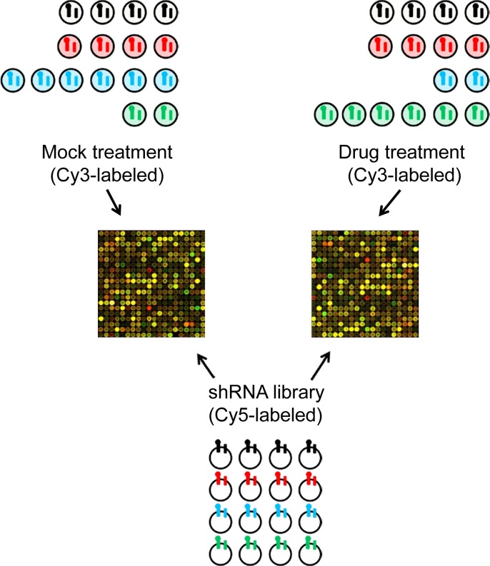
Figure 2. Schematic of the array protocol followed to readout the relative abundance of individual shRNAs following treatment.
The barcoded shRNA inserts from mock or drug treated cells are labeled with Cy3-nucleotides and used as probes in a competitive two-color hybridization with Cy5-labeled probe generated from the barcoded shRNA inserts amplified from the E. coli generated library.

