
Figure 7.
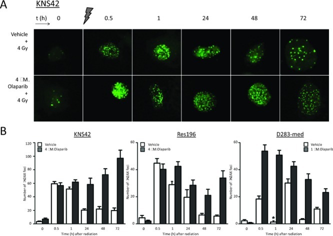
(A) Representative examples of immunofluorescent staining of γH2AX foci before, and (0.5 – 72 hours) after 4 Gy gamma radiation in KNS42 cells, pretreated with vehicle or 4 μM Olaparib. (B) Mean ± SE counts of γH2AX foci in KNS42, Res196 and D283-cells. D283-med cells were pretreated with 1 μM Olaparib. One hour after radiation nuclei stained diffusely positive for γH2AX in D283-med cells (*), though few foci could be detected.