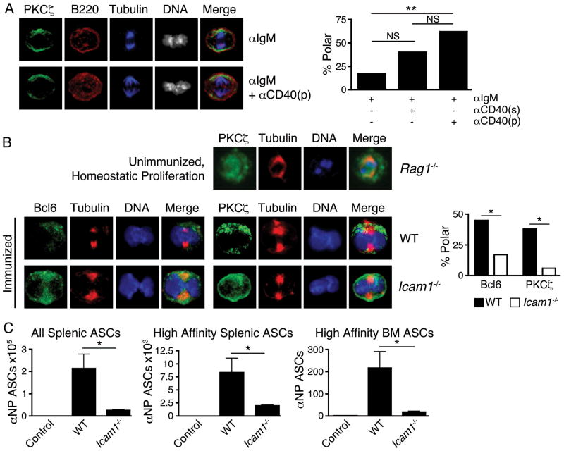
Fig. 2.
Polarity cues are required for asymmetric division of B cells. (A) Naive B cells simulated 36h with anti-IgM, with or without anti-CD40 to mimic a key contact-mediated signal from TFH cells. Anti-CD40 was withheld, added solubly to media (“s”), or coated on cell culture plates (“p”). Left, representative microscopy of no added anti-CD40 (upper) and plate-bound anti-CD40 (lower). Right, quantification of mitoses exhibiting asymmetry of PKCζ in indicated conditions. Left-to-right, n=18, 20, 21 mitoses, respectively. (B) Upper row, B cells undergoing homeostatic proliferation in Rag1−/− mice exhibit low incidence of asymmetric mitoses (11%, n=19 mitoses from pooled spleens of 3 recipients) compared to GC B cells from immunized mice (38%, n=16 mitoses); p<0.05. Lower rows, defective asymmetric division in adhesion-deficient mice. Left, GC B cells from 3 pooled spleens of d8 immunized wild-type (“WT”) and Icam1−/− mice stained for indicated components. Right, quantification of GC B cell mitoses exhibiting asymmetry of Bcl6 and PKCζ from indicated genotypes. Left-to-right, n=33, 18, 16, 18 mitoses, respectively. NS = p>0.05, * = p<0.05, ** = p<0.01 by chi-squared test. Similar results obtained at d5. (C) Icam1−/− mice have impaired antibody responses to immunization. Quantification of antibody secreting cells (ASCs) from immunized WT and Icam1−/− mice d14 post-immunization, compared to adjuvant-only injected WT mice (“Control”). n=6 mice/group, NS = p>0.05; * = p<0.05 by unpaired student’s t-test. All data are representative of 2 or more experiments.

