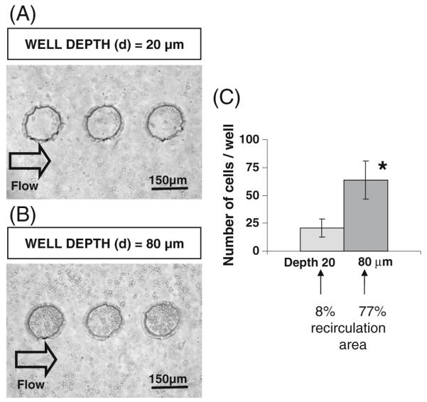
Fig. 5.

Fibroblast cells docked within microwells inside microfluidic devices. (a) Phase contrast image of cells in microwells with 150 μm in diameter and 20 μm in depth. (b) Phase contrast image of cells in microwells with 150 μm in diameter and 80 μm in depth. (c) Quantitative analysis of cell numbers docked inside microwells. Cell docking was significantly higher (p<0.01) in 80 μm thick microwells as compared to 20 μm thick microwells
