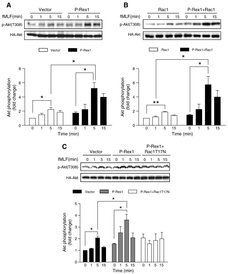
Fig. 11.
Effects of P-Rex1 and Rac1 on Akt phosphorylation. COSphox cells were transfected with expression constructs coding for FPR, P-Rex1 and, in some samples, Rac1 WT or Rac1 T17N. A HA-tagged Akt1 WT construct was co-transfected in all samples. Phosphorylation of Akt1 was determined using an anti-phospho-Akt (Thr308) antibody, in fMLF (1 μM) stimulated cells expressing control (vector), P-Rex1 (A), P-Rex1 plus Rac1 (B), and P-Rex1 plus Rac1 T17N (C). Total HA-Akt1 in the same cell lysate was determined. The relative level of Akt phosphorylation was shown in bar graphs below each blot. Densitometric analysis was performed and relative phosphorylation of Akt was determined after normalization against the total Akt level in transfected cells. Data were represented as mean±SEM of three independent experiments. * P < 0.05, ** P < 0.01.

