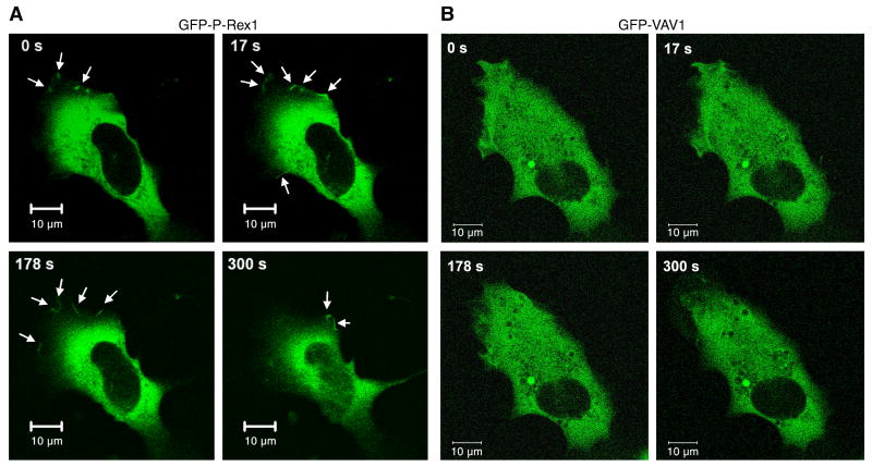
Fig. 2.
P-Rex1 membrane localization in reconstituted COSphox cells. COSphox cells were transfected with an expression vector for FPR and either the EGFP-P-Rex1 construct (A) or EGFP-Vav1 construct (B) or EGFP vector without cDNA insert (not shown). For each group, images of the same cells were taken at different time points after fMLF stimulation. Arrows indicate the appearance of EGFP fluorescence. Data shown are representative of 3 independent experiments on different days. Scale bar = 10 μm. A colored version of this figure is available online.

