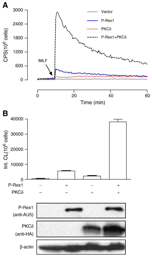
Fig. 7.
PKCδ potentiates P-Rex1-dependent superoxide generation. The fMLF-induced superoxide generation was determined in COSphox cells expressing FPR together with Prex1, PKCδ or both. (A) Production of superoxide as determined using isoluminol-enhanced chemiluminescence in fMLF (1 μM) stimulated cells. (B) Quantification of superoxide produced in the first 20 min after fMLF stimulation, based on integrated chemiluminescence (Int. CL). The relative expression levels of the transfected constructs are shown below the bar graph. At least 3 experiments were performed, and similar results were obtained. Representative data are shown. A colored version of this figure is available online.

