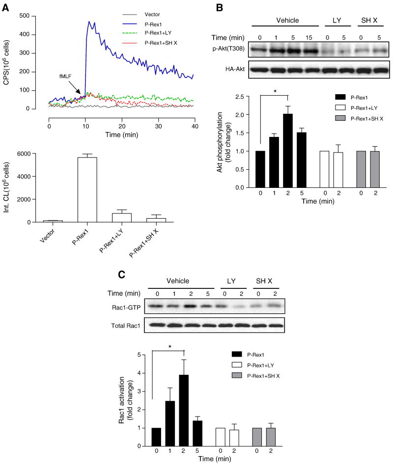
Fig. 8.
Regulation of P-Rex1-mediated superoxide generation and Rac1 activation by PI3K and Akt. Transfected COSphox cells expressing FPR and P-Rex1 were treated with the PI3K inhibitor LY294002 (50 μM) or the Akt inhibitor SH X (5 μM) for 5 min before fMLF (1 μM) stimulation. Quantification of the data was shown in bar graph below, based on integration of chemiluminescence in the first 20 min after fMLF stimulation. (B) The transfected COSphox cells were similarly treated with LY294002 or SH X as in (A), and fMLF-induced Akt phosphorylation was determined with an anti-pAkt (Thr308) Ab. Densitometric analysis was performed and relative phosphorylation of Akt was determined after normalization against total Akt in the same sample. Data shown are mean±SEM and are representative of three independent experiments. * P<0.05. (C) fMLF-induced Rac1 activation was determined using RBD-GST pull-down assay, in the transfected COSphox cells treated with either LY294002 or SH X treatment as in (A). Densitometric analysis was performed and relative level of activated Rac1 was determined after normalization against total Rac1 in the same samples. Data shown are mean±SEM and are representative of three independent experiments. * P<0.05. A colored version of panel (A) is available online.

