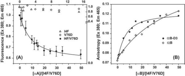
Figure 4.
Binding isotherms of γD-crystallin mutant to (A) αA-crystallin and (B) αB-crystallin and its phosphorylation mimic αB-D3. The concentration of the single mutants was 50 μM while the double mutant was 25 μM. Samples were incubated at 37 °C for two hours at the appropriate ratio as described in the methods section. The solid lines for the double mutant I4F/V76D in (A) and (B) are non-linear least-squares fit assuming high affinity binding where 4 α-crystallin subunits bind one γ-crystallin. The dissociation constants are 43±7 and 27±6 μM for αA-crystallin and αB-D3, respectively. For αB-crystallin, the dissociation constant was larger than 300 μM.

