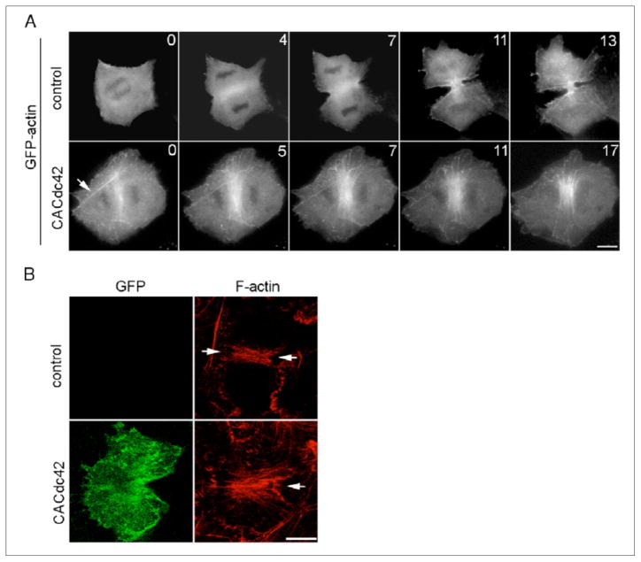
Figure 3. Effects of overexpression of constitutively active Cdc42 on equatorial actin assembly.
(A) Cells were transfected with GFP-actin (control) or both GFP-actin and mRFP-CACdc42 (CACdc42) and were monitored by time-lapse fluorescence optics. Thick actin bundles are observed outside the equator in cells transfected with mRFP-CACdc42 (arrow). Time elapsed in minutes since anaphase onset is shown at the top-right corner of each image. Scale bar represents 10 μm. (B) Cells transiently transfected with GFP-CACdc42 (green; GFP) or control non-transfected cells were stained with rhodamine-phalloidin (red; F-actin). Arrows indicate the position of the cleavage furrow. Three-dimensional reconstructed images are presented. The scale bar represents 10 μm.

