
Figure 4. Effects of depletion of Cdc42 and overexpression of constitutively active Cdc42 on RhoA localization during cytokinesis in NRK cells.
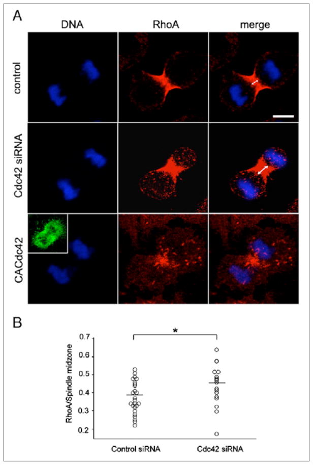
(A) Control non-transfected cells (control), cells depleted of Cdc42 (Cdc42 siRNA) and cells overexpressing GFP-CACdc42 (CACdc42) were fixed and stained for RhoA. The inset shows the image of GFP-CACdc42. Scale bar represents 10 μm. (B) The ratio of the width of the area stained with RhoA antibodies to that of the spindle midzone in control cells (n = 25; Control siRNA) and cells depleted of Cdc42 (n = 25; Cdc42 siRNA). *P < 0.05.