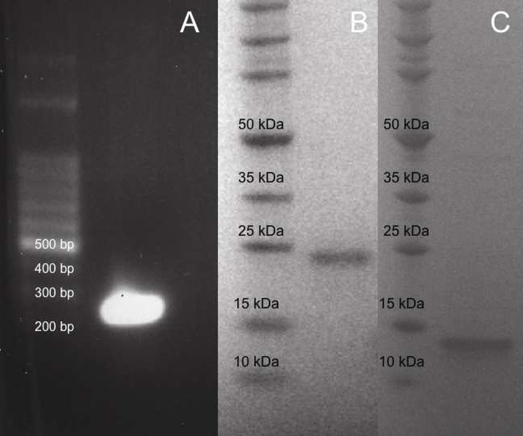
Fig. 1.
Cloning, expression, and purification of r-Rub. (A) 100 bp DNA marker (left lane). Rubistatin cDNA (right lane). SDS-PAGE of expressed protein resolved in a 4–12 % NuPAGE Bis-Tris gel under reducing condition (B–C). (B) Molecular weight marker (left lane). Thrx/r-Rub fusion protein (right lane). (C) Molecular weight marker (left lane). Purified r-Rub peptide (right lane).

