
Figure 1.
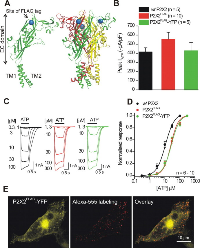
The FLAG tag at K79 in P2X2 receptors is surface exposed and can be labeled without altering receptor function. A, The models show a single P2X2 subunit (left) and a trimeric P2X2 receptor (right) with the FLAG tag insertion site shown schematically as a blue sphere (at K79) in the extracellular (EC) domain. The diagrams are based on the recently published zebrafish P2X4.1 crystal structure. B, Peak ATP-evoked current densities were not significantly different for the FLAG-tagged and FLAG plus YFP-tagged P2X2 receptors relative to WT P2X2. C, Representative traces for ATP-evoked currents at the indicated ATP concentrations for WT P2X2, P2X2FLAG, and P2X2FLAG–YFP receptors. D, Normalized ATP concentration–effect curves for WT P2X2, P2X2FLAG, and P2X2FLAG–YFP receptors (n = 6–10). E, Representative confocal stacks of a HEK-293 cell expressing P2X2FLAG–YFP receptors and labeled with anti-FLAG antibodies conjugated to Alexa Fluor 555, which labeled only the surface P2X2 receptors (representative of 3 such experiments).