Figure 3.
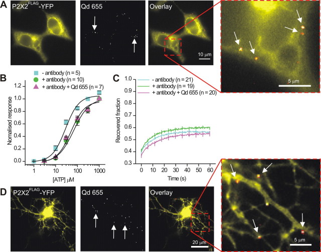
P2X2FLAG–YFP receptors can be labeled with Qds without markedly altering receptor function or macroscopic mobility. A, Labeling of P2X2FLAG–YFP receptors in HEK-293 cells. The panels show images of an HEK-293 cell expressing P2X2FLAG–YFP receptors. The YFP image (left) reveals overall P2X2FLAG–YFP expression in the cell, whereas the Qd image shows several spots of fluorescence (indicated by arrows). This is more readily seen in the merged image on the right. The boxed region for this image has also been enlarged. B, Concentration–effect curves for P2X2FLAG–YFP receptors expressed in HEK-293 cells under the indicated conditions. C, FRAP curves for P2X2FLAG–YFP receptors with (n = 20) and without (n = 21) Qd labeling. FRAP was performed and analyzed in 2 μm2 areas (see Table 1). D, As in A but for labeling of P2X2FLAG–YFP receptors in hippocampal neurons (the soma is overexposed to see the finer dendrites).

