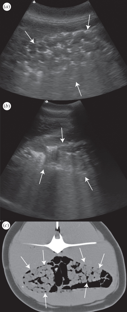
Figure 1.
Common dolphin IFAW10-069Dd. Arrows indicate renal margins. (a) B-mode ultrasound image of the left kidney showing hyperechogenicities (white) and ring-down artefact around the renules. (b) B-mode ultrasound image of the right kidney in the same dolphin with a greater depth of field to enhance the ring-down artefacts. (c) Transverse CT image at the level of the kidneys showing gas (black) surrounding and within the kidneys. The animal's left is to the left of the image. Gas is also seen in this image within intestinal loops and adjacent to the spinal cord. The CT was performed within 2 h of death. Window Width (WW) 553, Window Level (WL) 62. Three millimetre slice thickness, soft tissue reconstruction algorithm.

