
Figure 1.
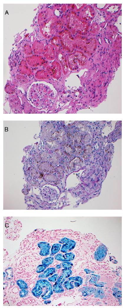
(A) Hematoxylin and eosin and (B) periodic acid–Schiff sections show brown pigment deposits in the proximal tubular epithelial cells accompanied by tubular degeneration and necrosis. (C) Prussian blue iron stain shows hemosiderin deposits in the tubular epithelial cells (A-C: original magnification, ×20).