
Fig. 2.
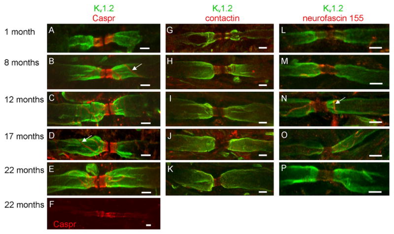
Paranodal protein domains are modestly altered in aged mice. In 1-month-old mice, Caspr (red) was restricted to the paranodal region (A). In 8- (B), 12- (C) and 17- (D) month-old mice, Caspr was observed outside the paranode; however, at these ages, extra-paranodal localization of Caspr was confined to the presumptive mesaxon (white arrows in B and D). In contrast, diffuse localization of Caspr was observed in juxtaparanodal and internodal regions of 22-month-old mice (F). In slight contrast with Caspr, contactin distribution was confined to the paranode (G–K). Similar to Caspr, neurofascin 155 was observed in all paranodal domains at all ages analyzed (L–P) and a line of immunoreactivity reminiscent of the presumed mesaxon labeled by the Caspr antibody was also observed in the anti-neurofascin labeled myelinated axons (white arrow in N). Note that Kv1.2 labeling (green) was occasionally observed in the paranode of young mice (G and L) but was confined to the juxtaparanode in adult and aged animals (H–K; M-P). Scale bars = 5 μm.