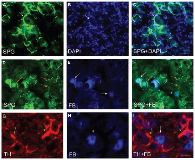
Figure 3.
Sympathetic sprouting surrounding the dorsal root ganglia (DRG) neurons. Histofluorescence staining for catecholamines in the thoracolumbar DRGs demonstrated thread-like sympathetic fibers (A, arrows) formed ‘basket’ structure surrounding the DRG neurons identified by DAPI staining (B). The sympathetic fibers extended between cells forming a cluster (C). The photomicrographs D–F demonstrated that SPG-stained fibers (green fibers indicated by orange arrows) were not surrounding but adjacent to FB-labeled colonic afferent neurons (blue cells indicated by white arrows). Tyrosine hydroxylase immunostaining of FB-labeled DRGs (G–I) also showed that sympathetic fibers sprouted not around but next to colonic afferent neurons. The photomicrographs showed examples from L1 DRG treated for tri-nitrobenzene sulfonic acid treatment for 7 days. Bars = 50 μm.

