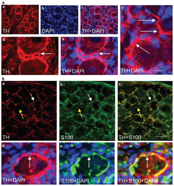
Figure 5.
Sprouting of sympathetic fibers around glial cells. Some of the tyrosine hydroxylase (TH) immunoreactive fibers (Aa) wrapped the satellite cells that were identified by nucleus staining with DAPI (Ab). In some cases, TH fibers extended from one cell to another (Ad, arrows), and sometimes a cluster of glial cells was wrapped together by TH fibers (Ae and Af, arrow). Double immunostaining showed that TH immunoreactivity was largely co-localized with the glial marker S-100 (Ba–Bc, white arrow indicated co-localization; yellow arrow showed S-100 fiber had no TH), and together these fibers surrounded the small satellite cells (Bd–Bf, arrow). The photomicrograph represented L2 dorsal root ganglia from animals sacrificed at day 7 post tri-nitrobenzene sulfonic acid treatment. Bar = 50 μm in Aa–Ac; 20 μm in Ad–Af; 60 μm in Ba–Bc; and 30 μm in Bd–Bf.

