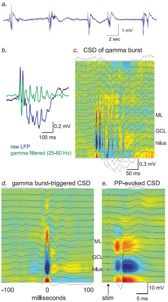
Figure 2. Characterization of spontaneous network activity in the dentate gyrus of urethane anesthetized mice.

a.) Raw LFP trace from a hilar recording electrode showing a series of spontaneous gamma bursts. Bursts occurred at an average of approximately 1 Hz in both groups (see Supp. Fig. 5). b.) Raw (blue) and gamma-filtered (green) LFP trace from a single gamma burst. The gamma-filtered trace was band-pass filtered at 25-80 Hz. c.) Current-source density analysis of the same gamma burst as in “B.”, over all 16 electrodes in the probe array, at 50 μm spacing. Current sinks, denoting influx of positive charge, are marked in red, while current sources are labeled in blue. d.) CSD of average gamma peak-triggered LFP from all gamma bursts from the experiment shown in panels b & c. e.) CSD of perforant-path evoked stimulation from the same experiment as panels b-d. Note that the pattern of sources and sinks during gamma bursts is generally similar to that evoked by perforant-path stimulation. Error bars are s.e.m. throughout.
